44 cell structure with labels
Cell Structure | SEER Training - National Cancer Institute Cell Structure. Ideas about cell structure have changed considerably over the years. Early biologists saw cells as simple membranous sacs containing fluid and a few floating particles. Today's biologists know that cells are infinitely more complex than this. There are many different types, sizes, and shapes of cells in the body. Cell: Structure and Functions (With Diagram) - Biology Discussion Eukaryotic Cells: 1. Eukaryotes are sophisticated cells with a well defined nucleus and cell organelles. 2. The cells are comparatively larger in size (10-100 μm). 3. Unicellular to multicellular in nature and evolved ~1 billion years ago. 4. The cell membrane is semipermeable and flexible. 5. These cells reproduce both asexually and sexually.
Plant Cell: Meaning, Components, Structure, Functions & Parts - Embibe Let us have a detailed look at the plant cell, its structure and the functions of different organelles. Components of a Plant Cell. The small membrane or non-membrane bound structures that are found in the cytoplasm or cellular matrix of a cell that works in a coordinated manner to maintain the homeostasis of a cell are termed as cell organelles.

Cell structure with labels
File:Plant cell structure svg labels.svg - Wikipedia File:Plant cell structure svg labels.svg. Size of this PNG preview of this SVG file: 649 × 475 pixels. Other resolutions: 320 × 234 pixels | 640 × 468 pixels | 1,024 × 749 pixels | 1,280 × 937 pixels | 2,560 × 1,874 pixels. This is a file from the Wikimedia Commons. Information from its description page there is shown below. A Labeled Diagram of the Animal Cell and its Organelles A Labeled Diagram of the Animal Cell and its Organelles There are two types of cells - Prokaryotic and Eucaryotic. Eukaryotic cells are larger, more complex, and have evolved more recently than prokaryotes. Where, prokaryotes are just bacteria and archaea, eukaryotes are literally everything else. › doi › fullEndothelial cell death after ionizing radiation does not ... Jul 18, 2022 · Here, we show that radiation induces endothelial cell death in tumors, yet the effects on vascular structure are minimized because the death occurs preferentially in small non-perfused vessels. On the one hand, vascular injury has been proposed as a prominent factor governing tumor response to radiation therapy.
Cell structure with labels. Labeled Plant Cell With Diagrams | Science Trends The parts of a plant cell include the cell wall, the cell membrane, the cytoskeleton or cytoplasm, the nucleus, the Golgi body, the mitochondria, the peroxisome's, the vacuoles, ribosomes, and the endoplasmic reticulum. Parts Of A Plant Cell The Cell Wall Let's start from the outside and work our way inwards. › jking540 › cell-biology-pptCell Biology ppt - SlideShare Sep 16, 2014 · Chapter 4 Objectives Section 3 Cell Organelles and Features • Describe the structure and function of a cell’s plasma membrane. • Summarize the role of the nucleus. • List the major organelles found in the cytosol, and describe their roles. • Identify the characteristics of mitochondria. Bacterial Cell Structure Labeling Diagram | Quizlet Cell Membrane A thin sheet of lipid and protein that surrounds the cytoplasm and controls the flow of materials in and out of the cell S Layer Monolayer of protein used for protection and attachment Fimbrae Fine bristles extending from cell surface that help in adhesion to other cells and surfaces. Outer Membrane Label Cell Parts | Plant & Animal Cell Activity | StoryboardThat Have your students label a plant and animal cell using one of the landscape poster layouts (small or large). Students will create a cell diagram labeled with the different organelles of plant and animal cells. The cell diagrams are easily colorable, allowing students to differentiate the different parts of the plant and animal cell quickly.
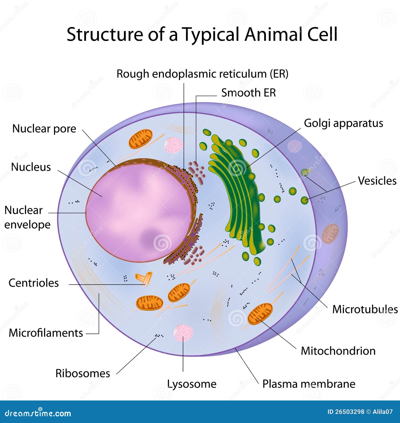
› learn › natural-scienceConstruction of the Cell Membrane - Wisc-Online OER Semi-permeable means "allowing certain substances to pass through it but not others, especially allowing the passage of a solvent but not of certain solutes." The cell membrane allows some things to pass through, and not others, which is why it needs transport proteins to let the other things through that the cell needs. Animal Cell Diagram with Label and Explanation: Cell Structure, Functions Animal cell is a typical Eukaryotic cell enclosed by a plasma membrane containing nucleus and organelles which lack cell walls, unlike all other Eukaryotic cells. The typical cell ranges in size between 1-100 micrometers. The lack of cell walls enabled the animal cells to develop a greater diversity of cell types. › en › productWhat's new in think-cell :: think-cell think-cell 8 greatly expands the slide layout functionality of our software by introducing the pentagon/chevron and textbox as new think-cell elements. Show project steps with accompanying bullet points by creating the basic structure very quickly out of building blocks, and use flexible single-click duplication to add additional steps. 03 Label the Cell Diagram | Quizlet Start studying 03 Label the Cell. Learn vocabulary, terms, and more with flashcards, games, and other study tools.
Animal Cells: Labelled Diagram, Definitions, and Structure - Research Tweet The endoplasmic reticulum (s) are organelles that create a network of membranes that transport substances around the cell. They have phospholipid bilayers. There are two types of ER: the rough ER, and the smooth ER. The rough endoplasmic reticulum is rough because it has ribosomes (which is explained below) attached to it. en.wikipedia.org › wiki › History_of_cell_membraneHistory of cell membrane theory - Wikipedia This was the first time the bilayer structure had been universally assigned to all cell membranes as well as organelle membranes. Evolution of the membrane theory. The idea of a semipermeable membrane, a barrier that is permeable to solvent but impermeable to solute molecules was developed at about the same time. cell membrane labels 31 Cell Membrane Diagram To Label - Labels For You duundalleandern.blogspot.com strcture PPT - POST LAB: Plant & Animal CELLS Or PowerPoint Presentation, Free cell cells onion plant membrane cellulose nucleus 100x lab animal ppt powerpoint presentation inside 36 Label Plasma Membrane - Labels 2021 documentdowu.blogspot.com plasma Plant and Animal Cell: Labeled Diagram, Structure, Function - Embibe Double membrane-bound structures found only in the plant cells. 2. This is an autonomous organelle. 3. There are stroma or matrix and grana or stacked discs that are involved in photosynthesis. 4. Grana are the site for photochemical reactions of photosynthesis, while stroma is the site for biochemical reactions of photosynthesis.
Types of Cells Video Teaching Resources | ClickView
Cell - Label | Cell Structure Quiz - Quizizz 16 Questions Show answers Question 1 30 seconds Q. Label #3 answer choices Nucleus Mitochondria Vacuole Golgi Body Question 2 30 seconds Q. Label #4 answer choices Cell wall Cell membrane Nuclear membrane Gatekeeper Question 3 30 seconds Q. Label #5 answer choices Nucleus Nucleolus Endoplasmic Reticulum Mitochondria Question 4 30 seconds

Bio in the news: Signal Proteins for Plant Stem Cell Discovered
Cell Structure And Function | Science Trends Daniel Nelson. The cell structure is defined by the cell membrane, the cytoplasm, and the nucleus. A cell is the smallest unit of life and its structure helps it to work as the basic building block of biology. The cell function is to keep all of the functions of the body performing as intended. This includes keeping toxins out of the body, help ...

Cells, major tissue types , Epithelial Cells Flashcards | Easy Notecards
Plant Cell- Definition, Structure, Parts, Functions, Labeled Diagram The central vacuoles are found in the cytoplasmic layer of cells of a variety of different organisms, but larger in the plant cells. Structure of plant cell vacuoles. These are large, vesicles filled with fluid, within the cytoplasm of a cell. It is made up of 30% fluid of the cell volume but can fill up to 90% of the cell's intracellular space.
Epithelia: The Histology Guide
Cell Organelles - Types, Structure and their Functions - BYJUS Ribosomes are found in the form of tiny particles in a large number of cells and are mainly composed of 2/3rd of RNA and 1/3rd of protein. They are named as the 70s (found in prokaryotes) or 80s (found in eukaryotes) The letter S refers to the density and the size, known as Svedberg's Unit. Both 70S and 80S ribosomes are composed of two subunits.

Animal And Plant Cells.
Learn the parts of a cell with diagrams and cell quizzes Two major regions can be found in a cell. The first is the cell nucleus, which houses DNA in the form of chromosomes. The second is the cytoplasm, a thick solution mainly comprised of water, salts, and proteins. The parts of a eukaryotic cell responsible for maintaining cell homeostasis, known as organelles, are located within the cytoplasm.
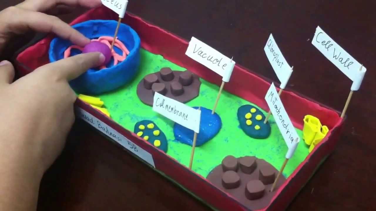
5th grade plant cell project - YouTube
cell membrane diagram with labels cell parts label animal plant membrane worksheet cells labeled structure labels functions worksheets diagrams visit. Name The Animal Cell Membrane : Cell Organelles | Plant Cell Vs. Animal michalleishere02949.blogspot.com. membrane sliderbase membranes pmf organelles ias. 30 Label Parts Of Cell Membrane - Labels Design Ideas 2020 ambitiousmares ...

Alila Medical Media | A typical cell, labeled diagram. | Medical illustration
Microscope Imaging Station. Gallery. - Exploratorium Elodea leaf cells with structures labeled . Chloroplasts and mitochondria move within Elodea leaf cells; nuclei are also visible as clear, fried-egg-shaped structures. Elodea are common freshwater aquarium plants. An elodea leaf was mounted in pondwater between a slide and coverslip with a silicon spacer. Images were taken on an inverted ...

Cells and Cell Structure - Earth Site Education
Cell Organelles- Definition, Structure, Functions, Diagram - Microbe Notes In a plant cell, the cell wall is made up of cellulose, hemicellulose, and proteins while in a fungal cell, it is composed of chitin. A cell wall is multilayered with a middle lamina, a primary cell wall, and a secondary cell wall. The middle lamina contains polysaccharides that provide adhesion and allow binding of the cells to one another.

Blogos: Metabolism VIII - Mother Letters, Elementals and Cellular Structure
Plant Cell - Definition, Structure, Function, Diagram & Types - BYJUS It is a rigid layer which is composed of polysaccharides cellulose, pectin and hemicellulose. It is located outside the cell membrane. It also comprises glycoproteins and polymers such as lignin, cutin, or suberin. The primary function of the cell wall is to protect and provide structural support to the cell.
/cell_organelles-56a09af25f9b58eba4b202f5.jpg)
Learn About Organelles
› books › NBK26880Looking at the Structure of Cells in the Microscope ... A typical animal cell is 10–20 μm in diameter, which is about one-fifth the size of the smallest particle visible to the naked eye. It was not until good light microscopes became available in the early part of the nineteenth century that all plant and animal tissues were discovered to be aggregates of individual cells.
/2000px-Plant_cell_structure_svg_vacuole.svg-58a886443df78c345bf8d009.png)
Vacuole Organelle
Structure of Bacterial Cell (With Diagram) - Biology Discussion It is a tough and rigid structure of peptidoglycan with accessory specific materials (e.g. LPS, teichoic acid etc.) surrounding the bacterium like a shell and lies external to the cytoplasmic membrane. It is 10-25 nm in thickness. It gives shape to the cell. Nucleus: The single circular double-stranded chromosome is the bacterial genome.

Cell Labeling - Simple and Complex
Chromosome Structure (Labeling) - The Biology Corner Chromosome Structure. Shannan Muskopf June 3, 2019. This simple worksheet shows a diagram of a chromosome and where it is located in the nucleus of the cell. Students use a word bank to label the chromatid, centromere, chromosomes, cell membrane, DNA, and nucleus. This worksheet was created for introductory biology for students to practice ...
A typical cell, labeled stock vector. Illustration of eukaryote - 26503298
Bacteria Cell Structures with labels - Dreamstime Bacteria Cell Structures with labels Royalty-Free Vector Bacterial cell structures labeled on a bacillus cell with nucleoid DNA and ribosomes. External structures include the capsule, pili, and flagellum. Morphology of internal structures of bacteria. cell anatomy bacteria, prokaryotic cell, cell, internal structures, prokaryotic, dna, bacteria,

EPS Vector - A typical cell, labeled, eps10. Stock Clipart Illustration gg62238928 - GoGraph
Cell Structure | Thermo Fisher Scientific - US Cell Structure. Organelle structures play a critical role in cellular function, and the detection of specific cell structures is key in fluorescence imaging of cells and tissue. Cell structure labels can be a counterstain method to identify the location of specific proteins and targets of interest within the cell.

34 Label The Cell Diagram - Labels Database 2020
Cell Structure and Function | General Biology Lab Manual Using Climate ... Cell Membrane, Chloroplast, Contractile vacuole, cytoplasm, flagellum, nucleus, nucleolus, pseudopod, ribosome, stigma. Name the three main layers of the plant cell wall, and list the chemical components of each layer. Describe the general structure and function of the nucleus. Describe the basic structure and function of each of the following ...
A Typical Cell, Labeled Stock Photo 112395266 : Shutterstock
Bacteria in Microbiology - shapes, structure and diagram - Jotscroll The bacteria shapes, structure, and labeled diagrams are discussed below. Sizes The sizes of bacteria cells that can infect human beings range from 0.1 to 10 micrometers. Some larger types of bacteria such as the rickettsias, mycoplasmas, and chlamydias have similar sizes as the largest types of viruses, the poxviruses.

Alila Medical Media | A typical cell, labeled diagram. | Medical illustration
A Labeled Diagram of the Plant Cell and Functions of its Organelles A Labeled Plant Cell Amyloplasts A major component of plants that are starchy in nature, the amyloplasts are organelles that store starch. They are classified as plastids, and are also known as starch grains. They are responsible for the conversion of starch into sugar, that gives energy to the starchy plants and tubers.
GitHub - madhubioinformatics/Enrichment: FatiGO. This is the conventional enrichment test where ...
| automated cell type annotation for scRNA-seq ... The programme uses cell-atlasing and cellular genetics to map cells in the human body combining cutting-edge methodologies and computational approaches. About CellTypist CellTypist was first developed as a platform for exploring tissue adaptation of cell types using scRNA-seq semi-automatic annotations.
Post a Comment for "44 cell structure with labels"