43 onion cells under microscope with labels
What organelles are in an onion cell? - Biology Stack Exchange To answer your question, onion cells (you usually use epithelial cells for this experiment) are 'normal' cells with all of the 'normal' organelles: nucleus, cytoplasm, cell wall and membrane, mitochondria, ribosomes, rough and smooth endoplasmic reticulum, centrioles, Golgi body and vacuoles. You cannot see most of these as they appear ... Onion Cell Lab Report.docx - Onion Cell Lab Report By - Course Hero Onion Cell Lab Report By : Nawaf Almalki Introduction: Many things that are viewed using a microscope, particularly cells, can appear quite transparent under the microscope. The internal parts of the cells, the organelles, are so transparent that they are often difficult to see. Biologists have developed a number of stains that help them see the cells and their organelles by adding color to ...
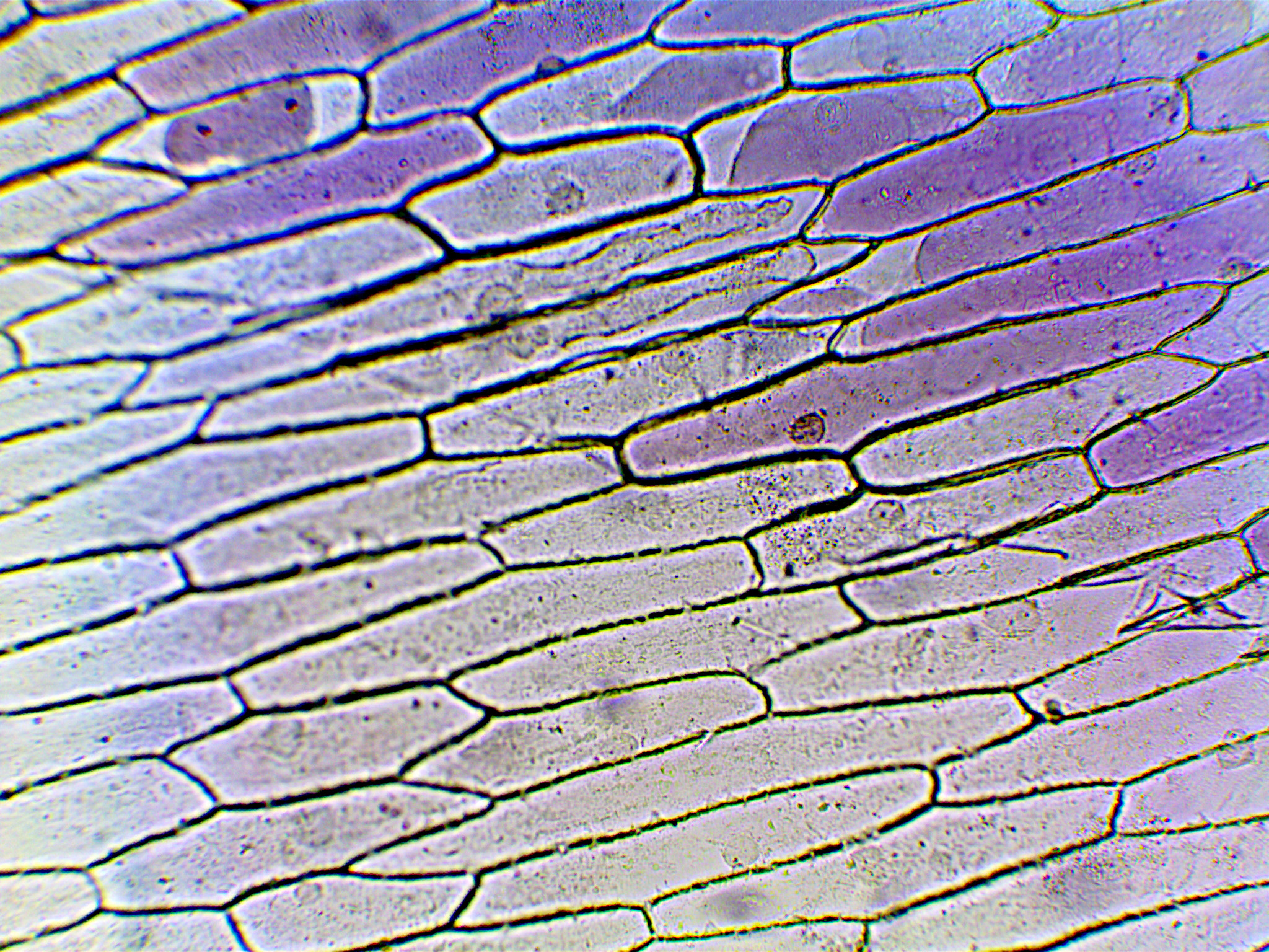
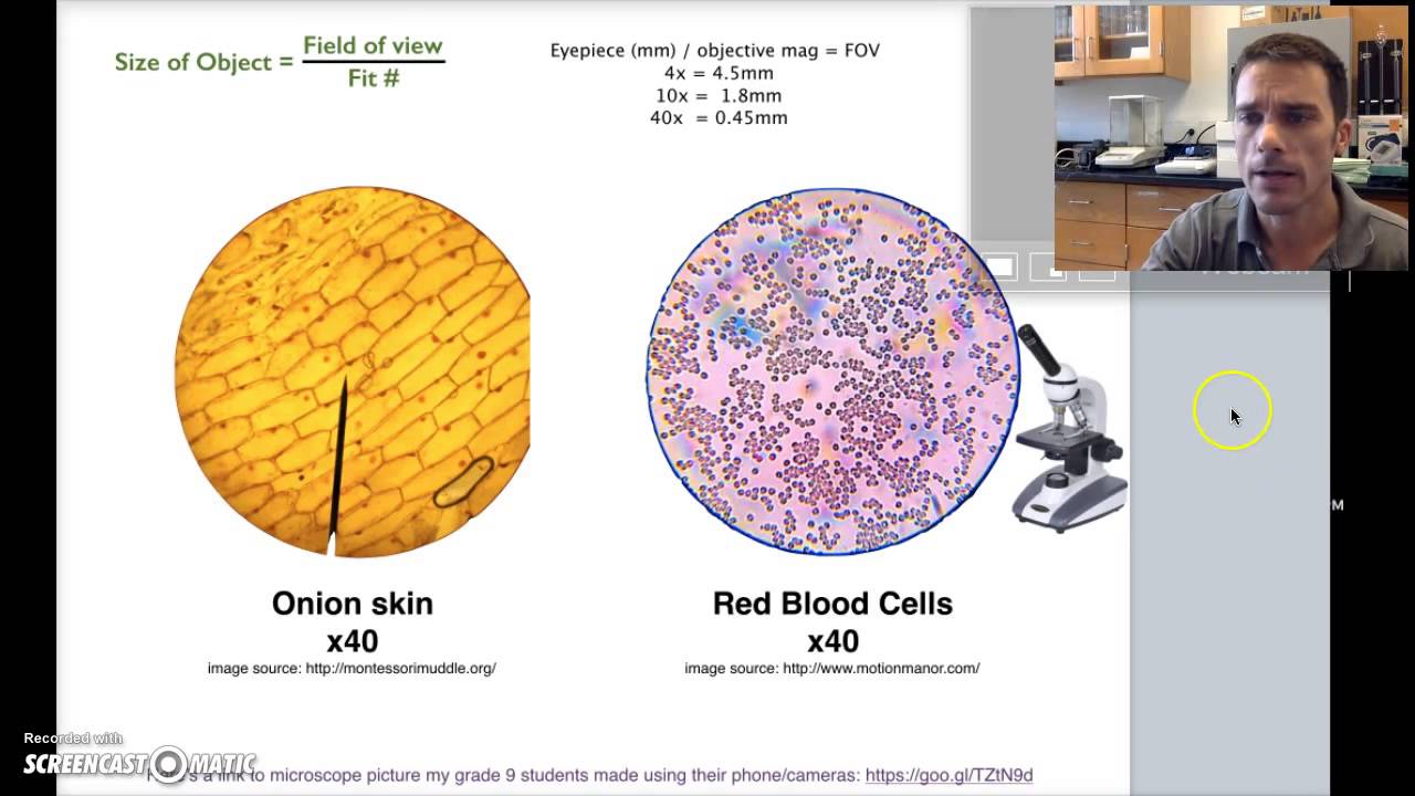
Using a light microscope - Required Practical Review Biology Practical - Using a light microscope to observe, draw and label cells in an onion skin. Video link: ...8 pages

Onion cells under microscope with labels
VIEWING PLANT CELLS UNDER THE MICROSCOPE: onion ... MICROSCOPE: onion cell preparation. This method allows students to view plant cells under the microscope. A single layer of onion.2 pages Onion Cells Under a Microscope (100x-2500x) - YouTube In this video you will see onion cells under a microscope (100x-2500x) as is, without any coloring. To observe the onion cells the thin membrane is used. It... Biology Project Cell Biology studying cells, mitosis, meiosis, the cell cycle, prokaryotes, eukaryotes, & viruses (en español - in italiano) Chemicals & Human Health basic toxicology, lung toxicology, environmental tobacco smoke & lung development, kidneys & metals
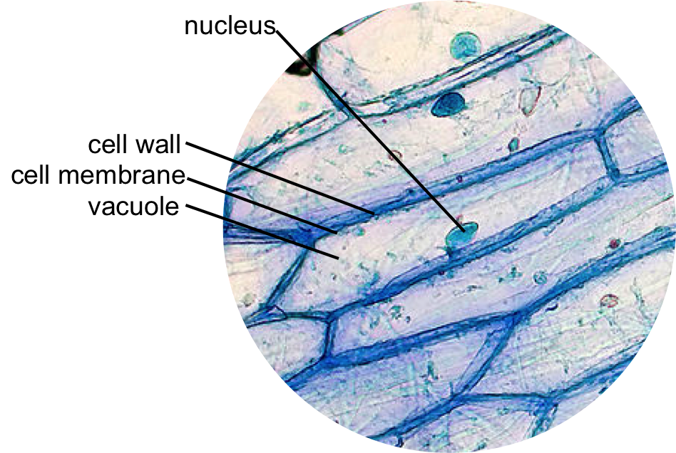
Onion cells under microscope with labels. Educational 01: To use a light microscope; 02: To obtain a good specimen of plant tissue for viewing under the microscope (onion cells) 03: To obtain a good specimen of animal tissue for viewing under the microscope (cheek cells) 04: To investigate the digestion of starch by amylase; 05: To investigate the effect of exercise on heart rate Under the Micrsocope: Onion Cell (100x - 400x) - YouTube In this "experiment" we will see onion cells under the microscope.For the experiment you will only need onion, dropper and the microscope (container and tool... Onion Cell Diagram Labeled Pdf ? - thesource2.metro Set your multimeter to measure current in the 20 mA range (the dial setting labeled "20m" on the right). Plug the multimeter's black probe into the port labeled COM. Plug the multimeter's red probe into the port labeled VΩmA. Use a red alligator clip lead to connect the multimeter's red probe to the positive (+) terminal of the 9 V battery. DOC The Onion Cell Lab - chsd.us Place the single layer of onion cell epithelium on a glass slide. Make sure that you do not fold it over or wrinkle it. Place a drop of iodine stain on your onion tissue. Put the cover slip on the stained tissue and gently tap out any air bubbles. Observe the cells under 4x, 10x, and 40x with the diaphragm wide open.
Microscope Cell Labeled Under Onion If you have a microscope (400x) and a properly stained slide of the onion root tip (or allium root tip), you can see the phases in different cells, frozen in time 1 millimeter long It is surrounded by cytoplasm Most of the cells will be parenchyma Find out how to observe cells under a microscope Property For Sale In National Parks Find out how ... Labeled Onion Cell Microscope Under (512) 585-1153 (cell) Kayak Committee Fred Wahlers cell 214-476-7725 [email protected] Complete a copy of Onion Cell Under Microscope Labeled (At minimum you should observe the nucleus, cell wall, and cytoplasm You will find collenchyma cells in dense clusters near the epidermis in a region called the cortex, forming the F showed you F showed ... Labeled Microscope Under Cell Onion Search: Onion Cell Under Microscope Labeled. Label the picture "Onion skin cells 400x" Values Equation Substitute Rearrange Answer Unit Identify and label the cell wall, cell membrane, nucleus, and nucleolus, if visible All bacteria are prokaryotes and stain with 18% alcoholic phloroglucinol-sulfuric acid solution and stain with 18% alcoholic phloroglucinol-sulfuric acid solution. PDF Onion Cells - Investigation - Exploring Nature 5. Observe the onion tissue under the microscope at 4x, 10x and 40x with lots of light (open diaphragm). Then slowly close the diaphragm while observing the image to find the best light for seeing cellular details. 6. Draw a section of onion skin cells at 10x magnification. Then switch to 40x and draw one cell and label it. Questions: 1.
Animal Cell Under Light Microscope Labelled : Draw and label the ... Students will observe onion cells under a microscope. We use microscope comprehensively in microbiology, mineralogy, cell biology, biotechnology, nano physics, microelectronics, pharmacology, and forensics. Magnification, however, is not the most important issue in microscopy. Observe the onion cell under both low and high power. ocr.org.uk › Images › 643844-question-paper-depth-inOxford Cambridge and RSA Friday 16 October 2020 – Morning 1 (a) A student was observing onion epithelial cells using a light microscope. They photographed these cells and the image obtained is shown in Fig. 1.1. The student then made a drawing of a few cells from this image. The drawing is shown in Fig. 1.2. Fig. 1.1 cytoplasm cell wall large permanent vacuole ribosome Fig. 1.2 Onion Skin Cells Labeled - the wonderful microworld onion skin cells ... Onion Skin Cells Labeled. Here are a number of highest rated Onion Skin Cells Labeled pictures on internet. We identified it from obedient source. Its submitted by executive in the best field. We... Cell Under Microscope Onion Labeled observe the onion under the low and high power of your microscope label the cell wall, cytoplasm, and the pigmented organelle structures (but you must label it with their real name using the evidence 1 look at each slide of specialised cells 04" (75mmx25mmx1mm) manufactured under iso 9001 quality control standard the diagram below shows onion …
Onion Cell Under Microscope - Personal Experience with Microscopes - AyushiSinhaMicroscopy ...
How to observe cells under a microscope - BBC Bitesize All living organisms are made up of cells. Cells are the smallest part of a living organism and are around 0.01 mm - 0.03 mm long. To look at a cell close up a microscope needs to be used.

onion cells through microscope | I put my camera right up to… | Flickr
sciencequiz.net › newjcscience › jcbiologyThe Cell - ScienceQuiz.net The diagram shows a group of onion cells. The parts labelled A, B and C respectively are ... The diagram shows a plant cell as seen under a microscope. Two of the ...

Phases of mitosis in control and 1-day CA-treated root tip cells. Bars... | Download Scientific ...
Labeled Under Cell Onion Microscope Observe under the microscope under low, medium and high powers Obtain a prepared slide of onion cells or prepare one yourself draw a cell of each of the required specimens and label correctly the parts seen under the microscope You will first observe the normal red onion cell and then see what happens when the environment around it is changed Post Lab Questions Post Lab Questions.

swifty science: onion cell lab
My onion cells at 40x magnification :) | Cellular level, Abstract, Neon ... This low magnification image shows an insular/organoid growth pattern and extensive areas of necrosis. Ramin, 13" x 20" Fall 2011, cuts on cotton paper, design derived from the cellular structures of South East Asian trees. Black Walnut, 13" x 20" Winter 2012, cuts on cotton paper, design derived from the cellular structure of the North ...
Microscope Onion Cell Labeled - Micropedia
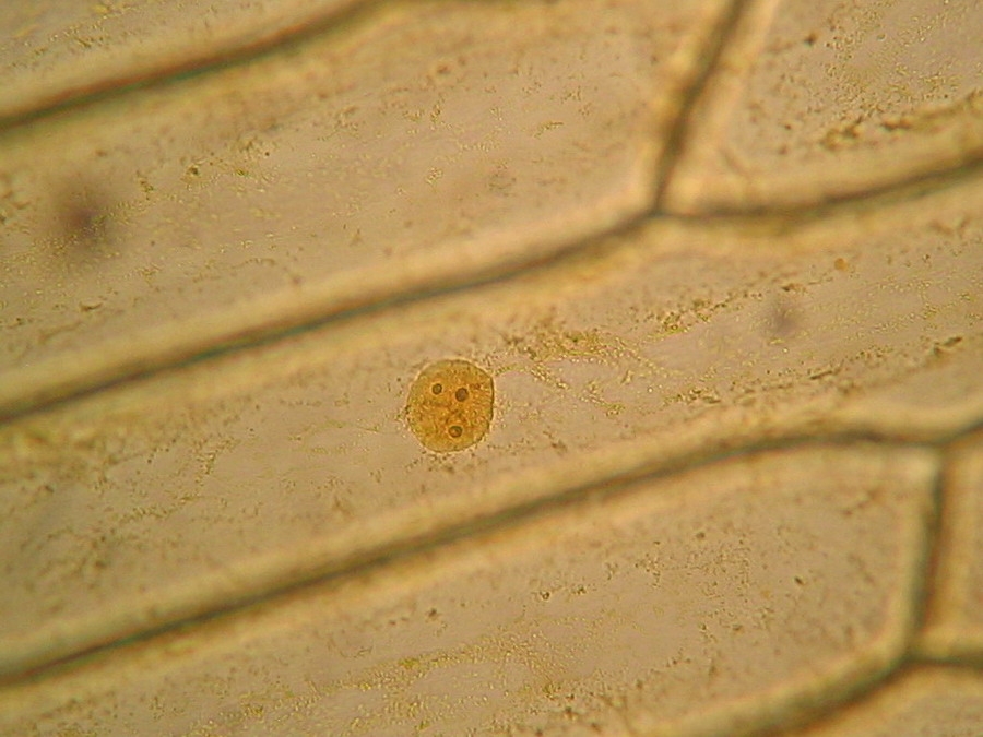
Onion Epidermis - kuensting.org Onion epidermal cells, iodine stain, 400X. The nucleus of an onion epidermal cell, 1000X magnification. ...
Labeled Onion Cell Under Microscope 40x - Micropedia
Plant Cell Under Microscope Labeled 40X : Young Root 2 Of Broad Bean ... Cells and viewing them under the microscope. A small square of a red onion skin (membrane) was observed under a microscope at high power (x40) magnification. (iv) describe how you applied the stain. They must draw and label the nucleus, cell membrane set up your microscope, place the onion root slide on the stage and focus on low (40x) power.
The inner epidermis of the onion bulb’s cataphylls (the onion skin).
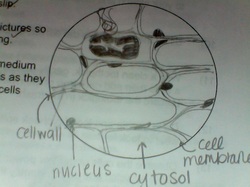
DOC Plant and Animal Cells Microscope Lab - Hillsboro City Schools Make a drawing of one onion cell, labeling all of its parts as you observe them. (At minimum you should observe the nucleus, cell wall, and cytoplasm.) Cheek cells 1. To view cheek cells, gently scrape the inside lining of your cheek with a toothpick. DO NOT GOUGE THE INSIDE OF YOUR CHEEK! (We will observe blood cells in a future lab!!) 2.

Living and Learning: Testing out my Microscope
Onion Cell - Weebly The cell of an onion peel consists of a cell wall, cell membrane, cytoplasm, nucleus and a large vacuole. The nucleus is present at the periphery of the cytoplasm. The vacuole is prominent and present at the centre of the cell. It is surrounded by cytoplasm.The vacuole is not visible under a microscope. Also, these cells have no chloroplasts as ...
The Cell — The Biology Primer
vii Sketch the onion peel cell as seen under the microscope Label the ... Put a drop of water in its centre and transfer the peel from the petridish to the slide with the help of a brush. Place the coverslip. (viii) Remove the extra water by placing the slide within a folded filter paper. (ix) Examine the slide first under low power and then under high power. (x) Record your observations.

Rens blog : Science, cells
Onion Root Tip Mitosis - Stages, Experiment and Results · Cover the sample (root tip) with a coverslip and gently press the coverslip down, then examine the slide under the microscope starting with low magnification * For this experiment, a properly prepared slide should appear light pink due to the stain to almost colorless. * Unused roots can be stored in 70 percent alcohol. Results

The Cells and Microorganisms Webquest
› 1814485 › Taiz_and_Zeiger_Plant(PDF) Taiz & Zeiger- Plant Physiology | Munish K Bansal ... Enter the email address you signed up with and we'll email you a reset link.
Onion Cells under Microscope
PDF Onion Cell Lab - somewaresinmaine.com Research Biology Onion Cell Lab page 1 of 3 Onion Cell Lab After you have completed the rest of this lab come back to this cover page DRAW & LABEL AN ONION CELL WITH ALL THE PARTS / ORGANELLES YOU OBSERVE UNDER 40X. Purpose: To observe and identify major plant cell structures and to relate the structure of the cell to its function. Materials: 1 ...

Onion Epidermal Cell Labeled - Top Label Maker
Onion Cells Under a Microscope - Requirements/Preparation/Observation Add a drop of iodine solution on the onion membrane (or methylene blue) Gently lay a microscopic cover slip on the membrane and press it down gently using a needle to remove air bubbles. Touch a blotting paper on one side of the slide to drain excess iodine/water solution, Place the slide on the microscope stage under low power to observe.
Onion Cell Under Microscope 4x 10x 40x - Micropedia
maths101.co.za › wp-content › uploadsNOVEMBER 2018 LIFE SCIENCES P1 - Maths 101 3.1 The root of an onion is a rapidly growing part of the onion. Many cells will be in different stages of mitosis. A sample of an onion tip was stained and studied under a microscope. The various phases of mitosis were identified and the number of cells counted in each phase. The results are recorded in the table below. Number of cells
Post a Comment for "43 onion cells under microscope with labels"